Hyaline Cartilage Thickness Measurement

How is the thickness of hyaline cartilage measured in a laboratory setting?

In a laboratory setting, the thickness of hyaline cartilage is typically measured using a specialized tool called a micrometer. This instrument allows researchers to accurately measure the thickness of the cartilage sample by gently pressing the micrometer against the tissue and recording the measurement. The thickness of hyaline cartilage is crucial in understanding its structural integrity and function within the body.

Inflammatory Response Evaluation

How is the thickness of hyaline cartilage measured in a laboratory setting?

What are the factors that can affect the accuracy of hyaline cartilage thickness measurements?

Several factors can affect the accuracy of hyaline cartilage thickness measurements. These factors include the angle at which the measurement is taken, the pressure applied during measurement, and the condition of the cartilage sample itself. It is essential for researchers to standardize their measurement techniques and account for these variables to ensure reliable and consistent results in their studies.

What to Expect in a Functional Capacity Evaluation

If an injury is keeping you from working, you may be wondering if and when you can return. It’s important to determine your current ability... The post What to Expect in a Functional Capacity Evaluation appeared first on BenchMark Physical Therapy.

Posted by on 2024-01-31

Can imaging techniques such as MRI or ultrasound be used to assess hyaline cartilage thickness?

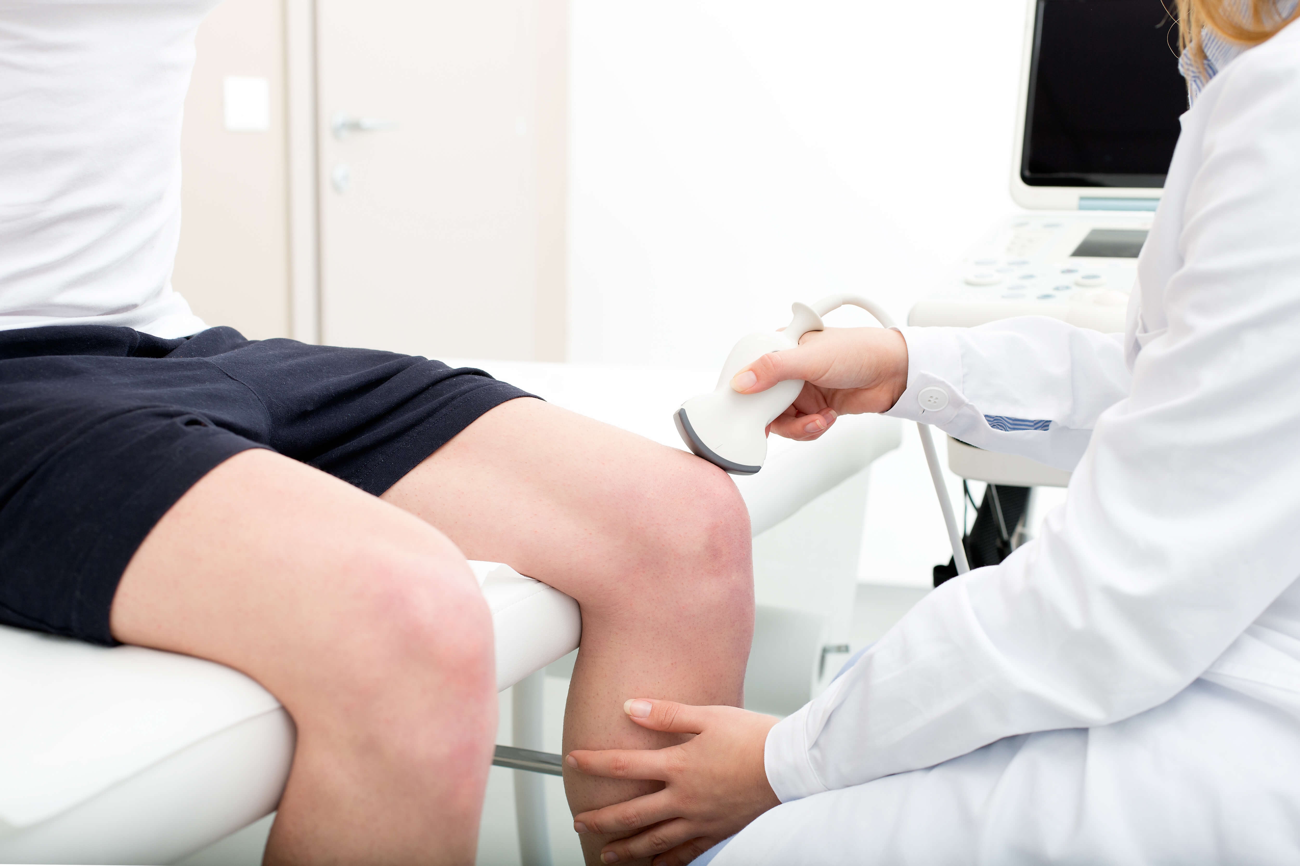
Imaging techniques such as MRI or ultrasound can indeed be used to assess hyaline cartilage thickness non-invasively. These imaging modalities provide detailed images of the cartilage structure, allowing researchers and healthcare professionals to measure the thickness of the cartilage in different joints of the body. MRI and ultrasound are valuable tools in diagnosing cartilage-related conditions and monitoring changes in cartilage thickness over time.

Can imaging techniques such as MRI or ultrasound be used to assess hyaline cartilage thickness?

Are there specific markers or stains that are commonly used to visualize hyaline cartilage in order to measure its thickness?

Specific markers or stains are commonly used to visualize hyaline cartilage in order to measure its thickness accurately. Safranin O, Alcian Blue, and Toluidine Blue are examples of stains that can highlight the cartilage matrix, making it easier to measure the thickness of the tissue under a microscope. These staining techniques are essential in histological studies of hyaline cartilage.

How does aging impact the thickness of hyaline cartilage in different joints of the body?

Aging can significantly impact the thickness of hyaline cartilage in different joints of the body. As individuals age, the cartilage may undergo degenerative changes, leading to a decrease in thickness and structural integrity. This age-related thinning of hyaline cartilage can contribute to joint pain, stiffness, and reduced mobility, highlighting the importance of monitoring cartilage thickness in older populations.

Soft Tissue Imaging As Utilized For Physical Therapy Rehabilitation

How does aging impact the thickness of hyaline cartilage in different joints of the body?
What role does nutrition play in maintaining the thickness of hyaline cartilage?

Nutrition plays a crucial role in maintaining the thickness of hyaline cartilage. Essential nutrients such as vitamin C, vitamin D, and omega-3 fatty acids are important for cartilage health and repair. A balanced diet rich in fruits, vegetables, lean proteins, and healthy fats can support the maintenance of hyaline cartilage thickness and overall joint health. Adequate hydration is also essential for cartilage function.

Are there any medical conditions or diseases that can lead to a decrease in hyaline cartilage thickness, and if so, how is this measured and monitored?

Several medical conditions or diseases can lead to a decrease in hyaline cartilage thickness, such as osteoarthritis, rheumatoid arthritis, and chondromalacia. These conditions can cause cartilage degeneration, resulting in a loss of thickness and integrity. Monitoring hyaline cartilage thickness in patients with these conditions is crucial for assessing disease progression, evaluating treatment effectiveness, and guiding clinical management strategies. Regular imaging studies and histological analyses are often used to measure and monitor changes in cartilage thickness in individuals with cartilage-related disorders.

Are there any medical conditions or diseases that can lead to a decrease in hyaline cartilage thickness, and if so, how is this measured and monitored?

Soft tissue imaging, such as ultrasound or MRI, plays a crucial role in diagnosing muscle tears during physical therapy by providing detailed visualization of the affected area. These imaging techniques allow physical therapists to accurately assess the extent and location of the tear, as well as identify any associated damage to surrounding structures. By examining the images, therapists can determine the appropriate treatment plan, including specific exercises, modalities, and interventions to promote healing and prevent further injury. Additionally, soft tissue imaging helps track the progress of the healing process, allowing therapists to make adjustments to the treatment plan as needed. Overall, the use of soft tissue imaging in diagnosing muscle tears enhances the precision and effectiveness of physical therapy interventions, leading to improved outcomes for patients.

Imaging techniques such as magnetic resonance imaging (MRI) and ultrasound can be utilized to detect signs of muscle compartment syndrome in rehabilitation patients. These imaging modalities can help identify increased pressure within a muscle compartment, swelling, inflammation, and potential muscle damage. By visualizing the affected area, healthcare providers can assess the severity of the condition and determine the appropriate treatment plan. Additionally, imaging can aid in monitoring the progression of the syndrome during rehabilitation and guide adjustments to the therapy regimen. Overall, incorporating imaging into the assessment of muscle compartment syndrome in rehabilitation patients can provide valuable insights into the underlying pathology and facilitate more targeted and effective interventions.

Soft tissue imaging, such as ultrasound or MRI, plays a crucial role in guiding the prescription of orthotic devices in physical therapy by providing detailed information about the structure and function of muscles, tendons, ligaments, and other soft tissues. By visualizing the specific areas of injury or dysfunction, physical therapists can accurately assess the extent of tissue damage, identify any underlying issues, and determine the most appropriate orthotic device to support and protect the affected area. This imaging technology allows for a more personalized and targeted approach to orthotic prescription, ensuring that the device fits properly and effectively addresses the patient's unique needs. Additionally, soft tissue imaging can help track progress over time, allowing therapists to make adjustments to the orthotic device as needed to optimize rehabilitation outcomes. Overall, the use of soft tissue imaging in guiding orthotic prescription enhances the precision and effectiveness of physical therapy interventions, leading to improved patient outcomes and recovery.

Imaging modalities commonly used to detect inflammation in synovial membranes during physical therapy include magnetic resonance imaging (MRI), ultrasound, and arthroscopy. These modalities can help visualize changes in the synovial membrane, such as thickening, increased vascularity, and fluid accumulation, which are indicative of inflammation. MRI is particularly useful for assessing soft tissue structures and detecting synovitis, while ultrasound can provide real-time imaging of the synovial membrane and surrounding structures. Arthroscopy, a minimally invasive procedure, allows for direct visualization of the synovial membrane and can also be used for therapeutic interventions. Overall, these imaging modalities play a crucial role in guiding treatment decisions and monitoring the effectiveness of physical therapy interventions for synovial inflammation.

Specialized imaging protocols for assessing lymphatic drainage in soft tissue injuries may include techniques such as lymphoscintigraphy, magnetic resonance lymphangiography, and dynamic contrast-enhanced magnetic resonance imaging. These protocols aim to visualize the lymphatic system, identify any disruptions or blockages in lymphatic flow, and assess the overall function of lymphatic drainage in the affected area. By utilizing these advanced imaging modalities, healthcare providers can gain valuable insights into the extent of lymphatic involvement in soft tissue injuries, allowing for more targeted treatment strategies and improved patient outcomes. Additionally, incorporating quantitative analysis of lymphatic function and flow dynamics can provide a comprehensive evaluation of lymphatic drainage patterns, aiding in the development of personalized treatment plans for individuals with soft tissue injuries.

Imaging markers that indicate the presence of myofascial trigger points in physical therapy patients may include increased muscle tension, localized areas of muscle stiffness, and the presence of taut bands within the muscle tissue. Additionally, imaging studies such as ultrasound or MRI may reveal changes in muscle fiber density, altered muscle metabolism, and decreased blood flow to the affected areas. These markers can help physical therapists identify and target specific trigger points for treatment, such as manual therapy techniques, dry needling, or therapeutic exercises aimed at releasing tension and improving muscle function. By utilizing imaging markers to pinpoint the location and severity of myofascial trigger points, physical therapists can develop more effective treatment plans to alleviate pain and improve overall function in their patients.